Description
Features:
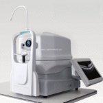
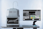
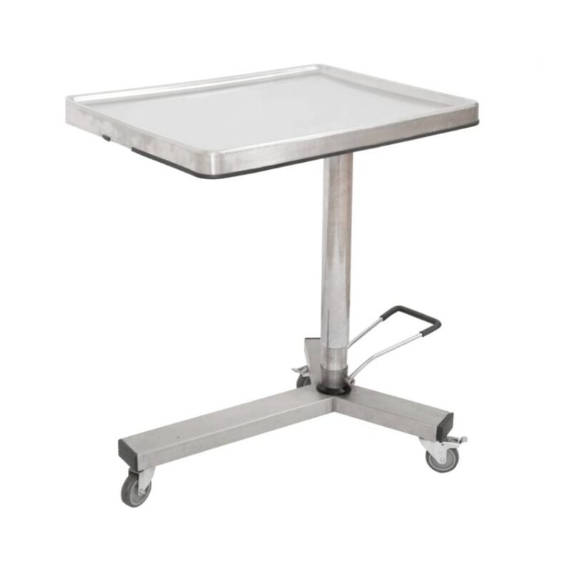
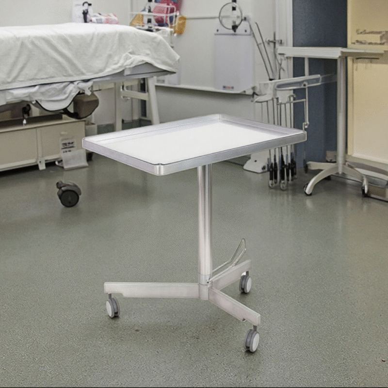
Compact Design Everything is inside this compact body. No external computer is needed.Plug in the power cable, then you areready to go.
PC inside Data acquisition and processing are accomplished by the internal computer. The data can be transported via ethernet or external harddrive. Peripheral devices, such as keyboard or printer, can be connected to the computer ports.
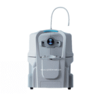
Easy Installation
No complex connection or setup.Compact body can fit in even smal!space.
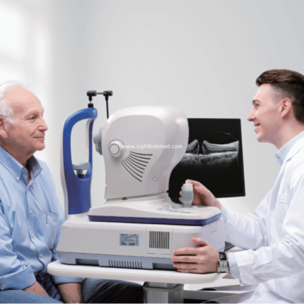
Simple Operation:
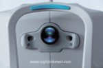
Step 1 Choose left or right eye. Push the direction buttons to move the target eye to the center of the field of view, then start data acquisition.
Step 2 Enter the data acquisition interface.The device is able to search for the OCT signal and optimize it automatically.
Step 3 Browse and analyze the acquired images.
| Methodology |
Spectral domain OCT |
| Axial resolution |
≤6 um (in tissue) |
| Transverse resolution |
≤20 um (in tissue) |
| Scan depth |
≥2.5 mm (in air) |
| Scan range |
≥6mm |
| Scan speed |
≥24,000 A-scans/sec, up to 36,000 A-scans/sec |
| Scan modes |
3D, Raster, Circle |
| Fundus image |
OCT en face |
| Focus adjustment |
-15D to +15D |
| Pupil diameter |
≥3mm |
| OCT light source |
840 nm SLD |
| Operation |
13.3″ touch screen, optional external mouse or keyboard |
| Power supply |
100-240 V, 50/60 Hz |
| Dimensions |
497 mm X 395 mm X 490 mm (LXWXH) |
| Weight |
34 kg (75 Ibs) |
| Analysis mode |
Up to 7 retinal layers segmentation, Macular analysis mode, RNFL & Optic Disk analysis mode, Glaucoma analysis mode and Progress analysis for follow-up examination. |
")
")
")
")