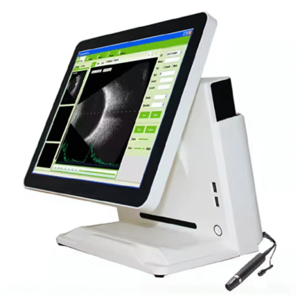
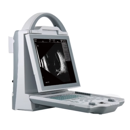
B scan:
Frequency: 10MHz/20MHz (optional) ,Magnetic driven, noiseless
Scanning Mode: Sector Scanning
Magnify:Multi continuous magnification,Real-Time magnification
Resolution: Lateral ≤0.3mm;Vertical≤0.2mm
Geometry position precision: Lateral ≤10%;Vertical≤5%
Depth:60mm
Enhance the part of vitreous body and retina
Gain of probe:30dB-105dB
Scanning Angle:53°
Gray Scale: 256
False Color: Multi colors. OCT
measurement type: multigroup distances, perimeters and areas
Image postprocessing: multiple curves processing, Pseudo-color processing curve
Movies: 100 images movie review, AVI JPG format image output
A scan:
Frequency:10MHz, with LED
Depth: 40mm
Precision:±0.05 mm
Measurement: Anterior chamber depth, lens thickness, vitreous body length, total length and average
Eye mode: Phakic / Aphakic / Dense / Various IOL
IOL Formula: SRK-II, SRK-T, HOFFER-Q, HOLLADAY,BINKHORST-II, HAIGIS
Stat. Calculation: Average and standard deviation
Store: 10 Scanning results for each eye
Others:
Display Mode :B,B+B,B+A,A
Hint: preset keyword
Case Search:Multi-keywords
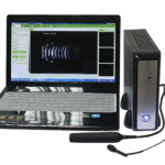
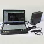
Working Platform: Windows XP,VISTA,WINDOWS7
User-defined report template poor credit secured loans