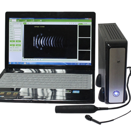
SL-500AB Ophthalmic Ultrasound A/B Scan
SL-500 A/B Scan with normal, vitreous body enhancement, retina observation mode, mainly used for diagnosis of intraocular diseases, display the location, shape range of the focus of infection and the relationship with the surrounding tissue.
Can be used for diagnosis on vitreous opacity, retinal detachment, eye base tumors etc. eye diseases. A scan is used to measure anterior chamber depth, lens thickness, axial length, calculate diopter of implant IOL as well.
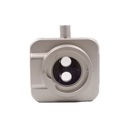
B Scan
Frequency: 10MHz/20MHz (optional) ,Magnetic driven, noiseless
Scanning Mode: Sector Scanning
Magnify:Multi continuous magnification,Real-Time magnification
Resolution: Lateral ≤0.3mm; Vertical≤0.2mm
Geometry position precision: Lateral ≤10%; Vertical≤5%
Depth: 60mm
Enhance the part of vitreous body and retina
Gain of probe:30dB-105dB
Scanning Angle: 53°
Gray Scale: 256
False Color: Multi colors. OCT
measurement type: multigroup distances, perimeters and areas
Image postprocessing: multiple curves processing, Pseudo-color processing curve
Movies: 100 images movie review, AVI JPG format image outputA scan:
Frequency: 10MHz, with LED
Depth: 40mm
Precision: ±0.05 mm
Measurement: Anterior chamber depth, lens thickness, vitreous body length, total length and average
Eye mode: Phakic / Aphakic / Dense / Various IOL
IOL Formula: SRK-II, SRK-T, HOFFER-Q, HOLLADAY,BINKHORST-II, HAIGIS
Stat. Calculation: Average and standard deviation
Store: 10 Scanning results for each eyeOthers:
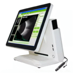
Display Mode: B, B+B, B+A, A
Hint: preset keyword
Case Search:Multi-keywords
Screen: 15 inch LCD
Built-in battery: can use 4 hours